Category: Case-Based Pearls

  • Case Example #2: Positional Saphenous and Posterior Tibial SSEP Changes During L2-4 Prone Transpsoas (PTP) Fusion

    Case Example #2: Positional Saphenous and Posterior Tibial SSEP Changes During L2-4 Prone Transpsoas (PTP) Fusion


    This case describes three separate interventions for positional lower extremity SSEP changes over the course of the first half of a L2-4 PTP with varying degrees of improvement in SSEP data. Interestingly, the evoked potential of most concern (left saphenous) remained the most stable compared to other waveforms throughout the procedure.

    Most of these changes occur either before retractors are placed, or on the contralateral side of interbody placement (working from left side). We show that it is possible to develop SSEP changes via overtightening of pelvic hip pads + strap.


    Patient was a 77 year old male with a history of low back and left-sided leg pain into his knee. He is 180cm tall and weighs 103kg. He denied any focal weakness. He has a history of previous left knee arthritis and replacement, coronary artery disease, and bladder cancer. He has lumbar stenosis, spondylosis with radiculopathy, and lumbar spondylolisthesis at the L2-4 levels.


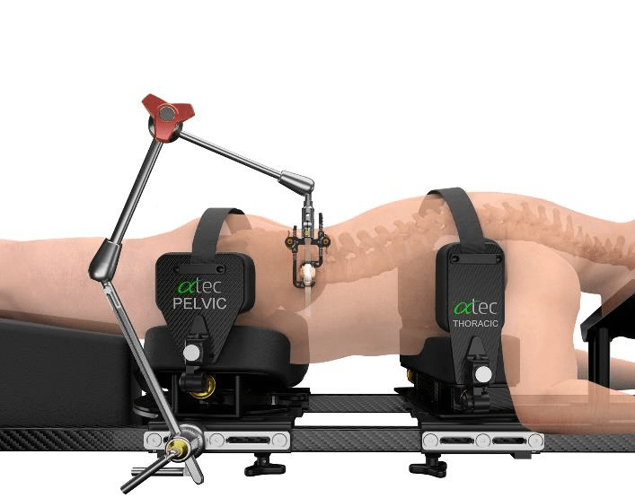
    Patient was positioned in the standard position for the prone transpsoas (PTP) approach (ATEC). Patient was turned on a Jackson table with chest and hip pads placed in the traditional fashion. Hip pads were secured with strap across buttocks.

    PTP Patient Positioner – ATEC Spine


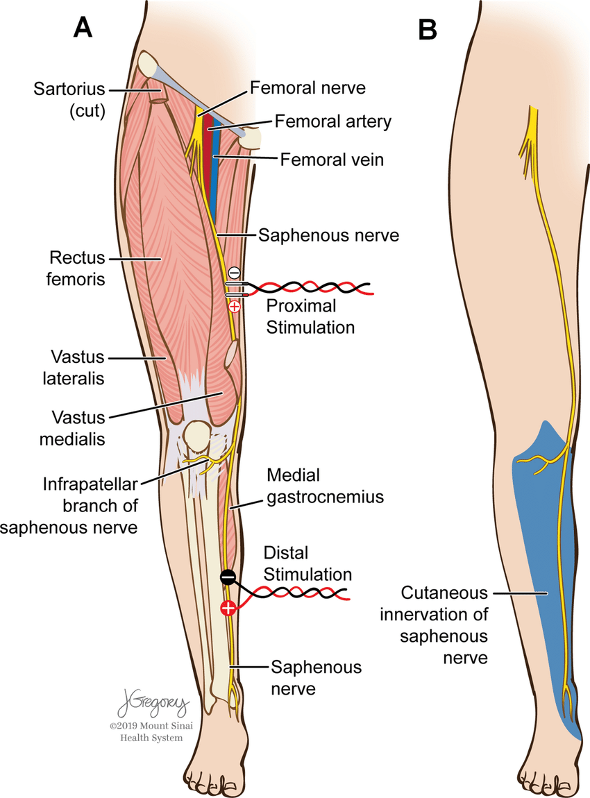
    Neuromonitoring included Somatosensory Evoked Potentials (SSEP) of the Posterior Tibial (standard ankle stimulation) and Saphenous (proximal- needle electrodes placed in thigh) Nerves and lower extremity electromyography (EMG), utilizing needle electrodes for vastus medialis & lateralis, biceps femoris, and anterior tibialis muscles (Standard ATEC SafeOp PTP Test). TOF was placed at the standard left peroneal nerve and was 4/4 throughout nerve & screw stimulation. All nerve stimulation trials were >10mA and all screws stimulated at >20mA.


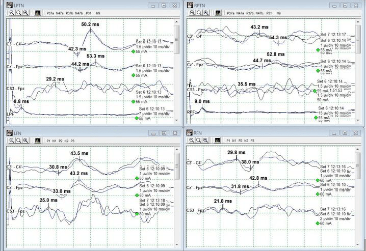
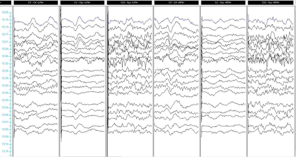
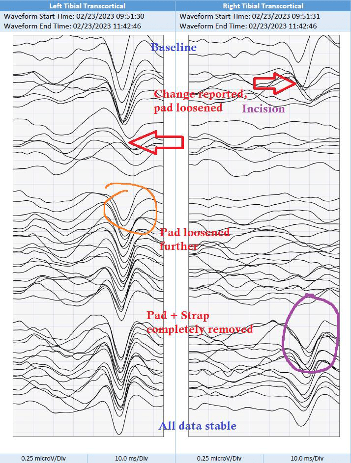
    Shortly after initial traces and baselines – and just around incision – a change in right tibial (Cortical + Transcortical) SSEPs were noted and reported (red arrows). Technical factors such as stimulation failure were ruled out as well as anesthetic factors, which had started at 1.5% and then 2.5% Sevofluorane during positioning, were mitigated by utilizing a total intravenous anesthetic (TIVA). MAP was maintained in the high 70s/low 80s. Propofol and remifentanil were maintained at 100mcg/kg/min and 0.10mcg/kg/min respectively throughout the remainder of the procedure. Left tibial SEPs and bilateral saphenous transcortical responses began to decrease in amplitude and demonstrate morphological changes while these changes were being discussed and troubleshooted.


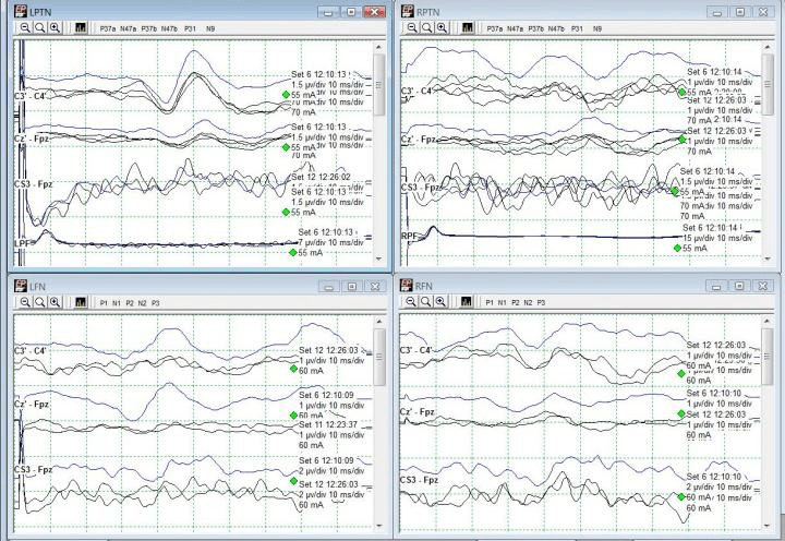
    The hip pad was not readily viewable due to c-arm telescope blocking, so it was unclear what could have been affecting positioning. Once c-arm was moved, the apparent right pelvic pad was loosened slightly to maintain current desirable positioning. A slight improvement in Right Saphenous and Left Tibial SEP was noted (orange circle) but further deterioration of bilateral Tibial SEPs persisted.


    Right tibial SEPs (Cortical + Transcortical) at this point were nearly abolished. The right pelvic pad was loosened further with moderate recovery of the bilateral saphenous SEPs (green circle).


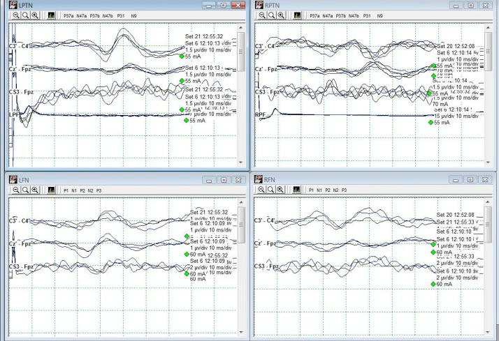
    Finally, the strap was cut and the pelvic pad was completely removed which allowed for a very rapid return of tibial cortical and transcortical responses (purple circle). These responses remained stable throughout the remainder of the procedure with no additional alerts. The patient went home the following day with no apparent deficit.


    Caution and care should be taken when placing and securing pelvic hip pads and straps. Improper positioning and overtightening of pads can lead to nerve fiber ischemia and peripheral nerve injury.

    Neuromonitoring must begin early and performed continuously throughout all surgical stages to identify changes in IONM data.

    Left & Right Saphenous Cortical SSEPs. Right Saphenous improvement circled in orange. Left saphenous demonstrates mild improvement after initial intervention.
    Left & Right Saphenous Transcortical responses. Improvements circled in green following second attempt at loosening pelvic padding.
    Left & Right Tibial Cortical responses. Changes from baseline noted with red arrow. Improvement circled in purple. Left improved with further loosening, right only returned after complete pelvic pad removal.
    Left & Right Tibial Transcortical responses. Changes from baseline noted with red arrow. Improvements circled in orange and purple. Left improved with further loosening, right only returned after complete pelvic pad removal.


    All data remained stable up and including to the end of monitoring.

  • Case Example #1 – Positional Saphenous Nerve SSEP Changes During L3-4 XLIF

    Case Example #1 – Positional Saphenous Nerve SSEP Changes During L3-4 XLIF

    The femoral nerve is the largest branch of the lumbar plexus. It originates from posterior L2 divisions to L4, and innervates the surfaces of the medial and anterior thigh. The saphenous nerve is monitored as the largest cutaneous branch of the femoral nerve to reflect femoral nerve sensory function during indicated procedures. For the lateral lumbar and eXtreme lateral interbody fusion (LLIF and XLIF) procedures, which are mostly performed between the L2-L5 levels, this main branch is mostly at risk by way of retraction systems in situ. This case presents an example of saphenous nerve SSEP changes with relatively stable tibial nerve SSEPs, further reinforcing the importance of saphenous nerve monitoring during lateral procedures.

    NuVasive XLIF Procedure Animation – NuVasive

    This patient was a 45-year old female, presenting with complaints of excruciating lower back and left-sided leg pain with associated numbness through her hip and into her foot. She was also beginning to experience these symptoms on the right. She had no weakness. She states she had no history of diabetes, hypertension, seizure, or stroke. Her prior surgical history included a lumbar fusion at L4-5 and a cervical fusion.

    She was positioned in the lateral decubitus position with her left side up and knees bent towards her chest. Tape was wrapped around the hips, just above the knee, and across the lower legs to keep her in place on the bed. Pillows were placed between the knees. Anesthetic protocol included a total intravenous method, utilizing between 100- 150mcg/kg/min of propofol and a steady 0.30mcg/kg/min of remifentanil. Surgeon requested SSEP monitoring (Saphenous + Posterior Tibial + Ulnar) with NuVasive EMG for dilator / nerve stimulation. Needle electrodes were placed in the standard placements for PTN, SN (Fig 1), & UN stimulation with additional needles behind the popliteal fossa and sticky pads over the femoral nerve as back-up for saphenous nerve.

    Figure 1: Stimulation placement for saphenous
    nerve SSEPs (figure from Silverstein et al. 2015)

    Baseline SSEPs were monitorable from all extremities except for RPTN, which was just barely marginally present. (Fig 2). Initial RPTN SSEPs were monitorable but quickly appeared to wash out following additional traces. PTN SSEPs were stimulated at 60mA 0.5ms 2.09Hz, SN SSEPs at 70mA 0.5ms 2.09Hz, and UN SSEPs at 30mA 0.3ms 2.09Hz. PTN + SN SSEPs were interleaved on the same timeline to more quickly acquire lower extremity data.

    Figure 2: Baseline PTN & SN SSEPs

    A change in the LSN SSEPs was reported almost immediately after setting baselines and prior to any implant sizing or placement (Fig 3). Some decompression work had begun but it was believed based on stimulation trials that the surgeon was well away from any significant nerve roots or groups. No problems in SSEP stimulation delivery were apparent based on movement during stimulation in all 4 extremities.

    The recommendation therefore was made to maintain an elevated blood pressure (declined due to surgeon preference for lower mean blood pressure for less bleeding) and to remove any distraction down at the legs. The surgeon declined this at the time as well, stating that he had a good deal of retraction in place and that it would interfere with surgical maneuvers. For the time being, the legs were only gently moved around and shaken to attempt to restore some blood flow to the legs.

    Figure 3: Loss of Left SN SSEP (Bottom Left)

    Immediately after instrumentation was in place, the tape across the hip, knee, and foot was cut away and removed. The left leg was lifted off the right and set back into its original position, only without tape. Within one data set, LSN SSEPs were within baseline parameters (from 90%+ reduced) and Right PTN SSEPs were much more reliable in cortical channels (Fig 4). Some improvement was seen in LSN subcortical responses as well.

    Figure 4: Recovery of Left SN SSEP after tape removal. Mild improvement also seen in R PTN SSEP (slightly more repeatable).

    The patient was then flipped to prone for posterior instrumentation with no significant events. She woke up with no additional deficits, and not much psoas pain which is very common following lateral spine surgeries. Because the actual data changes occurred in only LSN SSEPs, this case demonstrates the usefulness of saphenous and/or femoral nerve SSEPs in not only directly assessing L2-L4 nerve root function, but also as a reflection of possible malpositioning during lateral spine surgery.

    Monitoring PTN + SN/FN SSEPs on the same timeline can be helpful to evaluate most at-risk sensory pathways simultaneously. Positioning effects can occur prior to any monitoring and must therefore be approached quickly in the absence of monitorable baselines in patients with no pre-operative deficits.

    Figure 5: History stack of SN SSEPs illustrating loss + recovery. Change reported @ 12:24, Tape Removed @ 12:54.

    For more reading, please see the always-fantastic Dr. Silverstein’s article:

    Silverstein J, Mermelstein L, DeWal H, Basra S. Saphenous nerve somatosensory evoked potentials: a novel technique to monitor the femoral nerve during transpsoas lumbar lateral interbody fusion. Spine (Phila Pa 1976). 2014 Jul 1;39(15):1254-60. doi: 10.1097/BRS.0000000000000357. PMID: 24732850.